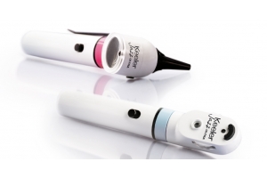
Although there are various types of ophthalmoscopes, two types typically come up in discussions: direct and indirect ophthalmoscopes.
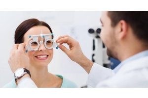
During an eye exam, eye physicians use an ophthalmoscope to examine the inside of the eye. This device sends a narrow, bright beam of light through the lens of the eye.
Ophthalmoscopes feature perforated lenses and mirrors that allow optometrists and ophthalmologists to detect diseases of the eye and other organs that may reflect in the eye.
In the next section, you will discover how direct and indirect ophthalmoscopes differ as well as the types of conditions this device can diagnose.
Direct Ophthalmoscope
The direct ophthalmoscope provides an upright, unreversed image of around 15 times magnification. A direct ophthalmoscope is a crucial tool for inspecting the fundus, which is the back portion of the eyeball. In most cases, it is advisable to conduct the examination in a darkened room.
As a physician, you should check the fundus for changes in pigment or color as well as changes in the caliber and shape of retinal blood vessels. Likewise, you should be on the lookout for abnormalities in the macula lutea, which receives and analyzes light from the center of the visual field.
Direct ophthalmoscopy also detects lens opacities and macular degeneration.
Explore our full direct ophthalmoscopes range here
What is an Indirect Ophthalmoscope?
An indirect ophthalmoscope produces a reversed or inverted direct image magnified two to five times.
When comparing direct vs indirect ophthalmoscopes, the indirect ophthalmoscope delivers a more powerful source of light. It also offers greater scope for stereoscopic inspection of the eyeball interior, and a specially designed objective lens.
The indirect ophthalmoscope has proven to be a valuable tool for treating and diagnosing detachments, holes, and tears of the retina.
For a successful examination, the patient's pupils must be completely dilated.
Binocular Indirect and Monocular Indirect Ophthalmoscopes
Indirect ophthalmoscopes can be divided into two different categories that include:
- Monocular indirect ophthalmoscopes
- Binocular indirect ophthalmoscopes
Monocular Indirect Ophthalmoscopes
Monocular indirect ophthalmoscopes offer a wider field of view and higher magnification levels than the traditional ophthalmoscope. As the name suggests, however, the monocular indirect ophthalmoscope only offers a single view of the eye's interior. For a physician to properly assess a patient's ocular condition and fundus, you should have the patient look in multiple directions.
Binocular Indirect Ophthalmoscopes
Instead of only projecting one, binocular indirect ophthalmoscopes project three elements in the eye. As a result, the ophthalmologist, or optometrist can get a three-dimensional rendition of the patient's interior eye, which facilitates a much more thorough examination.
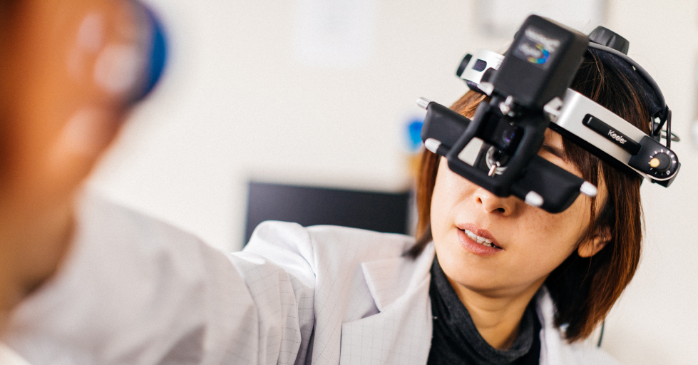
Indirect ophthalmoscopes are commonly mounted on the physician's head and can be used as a teaching aid. This device allows the lead practitioner to demonstrate diagnostic and procedural information to optometry students in the process of conducting an ocular exam.
Read more about our BIO range here
Direct vs Indirect Ophthalmoscope
While direct and indirect ophthalmoscopes are used to achieve similar goals, these devices have distinct differences. Use the chart below to see the differences between direct vs indirect ophthalmoscopes.
| Indirect Ophthalmoscopy | Direct Ophthalmoscopy | |
| Observation view field diameter | Wide view (approx. 37o in diameter) | Small view (approx. 10o in diameter) |
| Magnification | Five times when +13D condensing lense is used | Approx. 15 times |
| Structures viewed | Peripheral retina seen | Central retina only |
| Brightness | More brightness | Less brightness |
| Stereopsis | Binocular indirect provides superior stereopsis | Image created isn't stereoscopic |
| Image of fundus | Inverted and virtual image | Erect and real image |
| Scleral indentation | Easily achieved in binocular indirect ophthalmoscopy | Difficult |
| Retina anterior to the equator | More visual | Can be seen with difficulty |
| Visibility in hazy media | Better | Poor |
Looking Beyond the Eye with Ophthalmoscopes
These devices are integral in the examination of the patient's retina as well as the vitreous humor — the gelatinous liquid between the lens and retina. Even though the ophthalmoscope is a relatively simple, it can uncover a vast range of conditions, such as:
- Retinal detachment
- Eye tumors
- High blood pressure
- Glaucoma
- And more
The ophthalmoscope is typically used when patients complain of headaches and can be used to identify problems, such as swollen discs, arterial hypertension and intracranial pressure, which indicates more serious issues within the body.
In addition, ophthalmic professionals can identify "cupped" optic discs that are usually seen in patients with glaucoma. It can also be used to assess the ocular conditions of patients with diabetes to avoid vision loss related to diabetes or prevent diabetic retinopathy.
Contact Keeler
At Keeler Ophthalmic Instruments, we are industry leaders providing cutting-edge solutions and equipment. Whether you're considering a direct ophthalmoscope or an indirect ophthalmoscope, the experts at Keeler can help you find the best option for your practice.